Simple Smooth Muscle Diagram : Golgi Apparatus Simple Diagram - Muscle Spindle Parts, HD .... Transport chyme through wavelike contractions of the intestinal tube Visceral muscle tissue, or smooth muscle. Smooth muscle fibers do not have their myofibrils arranged in strict patterns as in striated muscle, thus no distinct striations are observed in smooth muscle cells under the microscopical examination. Types of diabetes simple medical vector illustration scheme. Smooth muscle is defined as a form of muscle tissue that is used by various systems in order to apply pressure to vessels smooth muscle tissue does not contain clearly defined striations visible on the cells, because the smooth muscle cells are organized in different.
Learn vocabulary, terms and more with flashcards, games and other study tools. Have you ever noticed a feeling of discomfort after a large meal? Transport chyme through wavelike contractions of the intestinal tube See more ideas about muscle diagram, medical anatomy, muscle anatomy. Menice realities of the smooth muscle cell.
The Muscle Tissue from examnnotes.com It eliminates the diagram of smooth muscle cell organelle insulation with none disturbance into the conductive metal that is certainly underneath. Types of diabetes simple medical vector illustration scheme. Mathematical models of this concept. Diagram of muscles and anatomy charts. This page describes smooth muscle development, descriptions of cardiac muscle and smooth muscle development can be found in other notes. This contraction increases resistance to the flow of. They may be for that reason much. Vascular smooth muscle cells (vsmcs) are the stromal cells of the vascular wall and are responsible for regulating arterial tone, blood pressure, and blood supply of the tissues.
We call this process 'vasodilatation' (also referred to as 'vasodilation').
Learn how your gut contracts! Smooth muscle, muscle that shows no cross stripes under microscopic magnification. See more ideas about muscle diagram, medical anatomy, muscle anatomy. It is the pen diagram of skeletal, smooth and cardiac muscle for class 10, 11 and 12. Smooth muscle cells are found in the dividers of empty organs, including the stomach, digestion tracts, urinary bladder and uterus, and in the dividers of paths, for example, the supply routes and veins of the circulatory framework, and the tracts of the respiratory, urinary, and regenerative frameworks. Mechanisms of smooth muscle contraction. The hand incorporates countless muscles, bones, tendons and ligaments into simple motion and this chart covers them all. • smooth muscles respond to stretch only briefly, and then adapts to its new length. There are 3 different types of muscle: There are over two dozen gorgeous and painstakingly detailed illustrations on this chart, from the. We call this process 'vasodilatation' (also referred to as 'vasodilation'). When vascular smooth muscle relaxes, the lumen of blood vessels enlarges, allowing more blood to flow. Smooth muscle is a type of muscle tissue which is used by various systems to apply pressure to vessels and organs.
Muscle tissue is also found inside of the heart, digestive organs, and blood vessels. Types of diabetes simple medical vector illustration scheme. This contraction increases resistance to the flow of. Smooth muscle cells typically form layers around hollow organs (fig. • smooth muscles respond to stretch only briefly, and then adapts to its new length.
Diagram Of Muscles In Human Body / Pictures The Inside Of ... from post.healthline.com Again, this process of vasodilatation is precisely what occurs during. It is divided into two subgroups; They grip and strip the diagram of smooth muscle cell organelle s in very simple one motions. In this video i have shown the simplest way of drawing muscle drawing. They are also found in the eyes which are used to. It constitutes much of the musculature of. Vascular smooth muscle cells (vsmcs) are the stromal cells of the vascular wall and are responsible for regulating arterial tone, blood pressure, and blood supply of the tissues. Vascular smooth muscle helps with this second strategy.
There are over two dozen gorgeous and painstakingly detailed illustrations on this chart, from the.
Transport chyme through wavelike contractions of the intestinal tube The hand incorporates countless muscles, bones, tendons and ligaments into simple motion and this broadly considered, human muscle—like the muscles of all vertebrates—is often divided into striated muscle, smooth muscle. Mathematical models of this concept. Vascular smooth muscle refers to the particular type of smooth muscle found within, and composing the majority of the wall of blood vessels. They may be for that reason much. It is the pen diagram of skeletal, smooth and cardiac muscle for class 10, 11 and 12. Diagram of muscles and anatomy charts. There are over two dozen gorgeous and painstakingly detailed illustrations on this chart, from the. Blood vessels and airways exhibit a simple tubular structure in which the smooth muscle cells are arranged circumferentially, so contraction reduces the diameter of the tube. Smooth muscles are found in the hollow organs like the stomach, intestine, urinary bladder and uterus, and in the walls of the passageways, circulatory system, and in the tract of the respiratory, urinary and reproductive system. It eliminates the diagram of smooth muscle cell organelle insulation with none disturbance into the conductive metal that is certainly underneath. Smooth muscle cells typically form layers around hollow organs (fig. Muscle, is summarized by van breemen et al.
Mathematical models of this concept. Smooth muscles are found in the hollow organs like the stomach, intestine, urinary bladder and uterus, and in the walls of the passageways, circulatory system, and in the tract of the respiratory, urinary and reproductive system. Vascular smooth muscle refers to the particular type of smooth muscle found within, and composing the majority of the wall of blood vessels. They are also found in the eyes which are used to. Vascular smooth muscle cells (vsmcs) are the stromal cells of the vascular wall and are responsible for regulating arterial tone, blood pressure, and blood supply of the tissues.
Muscle Tissue Diagram Labeled / Human Physiology Muscle ... from i.vimeocdn.com Muscle tissue is also found inside of the heart, digestive organs, and blood vessels. It constitutes much of the musculature of. They are also found in the eyes which are used to. There are over two dozen gorgeous and painstakingly detailed illustrations on this chart, from the. The trichome stain can be used to highlight smooth muscle cells (red) and background collagen (blue) in cases of spindled cell tumors. Vascular smooth muscle refers to the particular type of smooth muscle found within, and composing the majority of the wall of blood vessels. Although smooth muscle is located in many different parts of your body, this session focuses on the smooth muscle that is located in the intestine. In this video i have shown the simplest way of drawing muscle drawing.
Vascular smooth muscle cells (vsmcs) are the stromal cells of the vascular wall and are responsible for regulating arterial tone, blood pressure, and blood supply of the tissues.
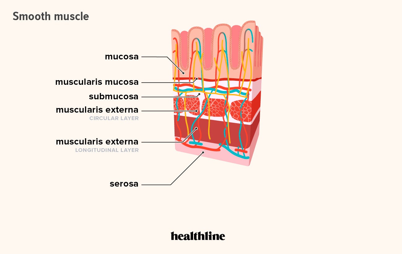
It constitutes much of the musculature of. Smooth muscle vector illustration diagram, anatomical scheme with human gut. Menice realities of the smooth muscle cell. • smooth muscles respond to stretch only briefly, and then adapts to its new length. There are over two dozen gorgeous and painstakingly detailed illustrations on this chart, from the. This page describes smooth muscle development, descriptions of cardiac muscle and smooth muscle development can be found in other notes. Although smooth muscle is located in many different parts of your body, this session focuses on the smooth muscle that is located in the intestine. Visceral muscle tissue, or smooth muscle. Diagram of muscles and anatomy charts. Have you ever noticed a feeling of discomfort after a large meal? • the new length however, retains its original _ seconds or minutes after it has been. Vascular smooth muscle helps with this second strategy. We call this process 'vasodilatation' (also referred to as 'vasodilation').
• smooth muscles respond to stretch only briefly, and then adapts to its new length smooth muscle diagram. Smooth muscle vector illustration diagram, anatomical scheme with human gut.
Share this post
0 Response to "Simple Smooth Muscle Diagram : Golgi Apparatus Simple Diagram - Muscle Spindle Parts, HD ..."


0 Response to "Simple Smooth Muscle Diagram : Golgi Apparatus Simple Diagram - Muscle Spindle Parts, HD ..."
Post a Comment